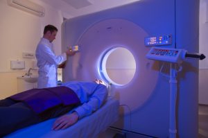
By using theragnostic radiotherapy, a personalized treatment plan can be tailored to both the geometric and biological attributes of a patient’s tumor.
Medical imaging needs radiation therapy just as radiotherapy needs imaging. Progress in functional and molecular imaging has dramatically improved the ability to map the spatial distribution of cellular phenotypes and microenvironmental parameters in 3D. Such maps may provide a stop-go switch for deciding whether to give a specific type of treatment.
By using radiation, it’s possible to exploit the full spatio-temporal information in a functional imaging map to titrate the biological effect in space and time. This would give physicians’ the ability to direct the radiation where it will be most affective in terms of the patient’s risk:benefit ratio.
This strategy for therapeutic use of imaging data is referred to as theragnostic imaging. Theragnostic radiotherapy involves the use of large amounts of functional imaging for treating the patient’s tumor growth.
 Conventional radiotherapy relies on accurate tumor detection, combined with optimal alignment of the radiation beam to the target, thus achieving successful treatments.
However, radiotherapy has been known to fail because of the high tumor burden, tumor hypoxia or tumor proliferation, for example. It has been shown that controlled dose escalation can overcome radiation resistance for multiple tumor types. It could even be possible to lower doses to radiosensitive tumors, thereby reducing the overall toxicity levels posed by radiation. In either case, theragnostics could play a major role.
The main theragnostic imaging modalities under investigation are positron emission tomography (PET) and magnetic resonance imaging (MRI), single-photon emission computerized tomography (SPECT), and magnetic resonance spectroscopy imaging (MRS/I).
According to Eric Paulson from Froedtert Hospital and the Medical College of Wisconsin, “Theragnostic radiotherapy is an emerging technique, still in its infancy. The hypothesis is that non-invasive biological images can be used to derive optimized, non-uniform prescription doses that can then be used in treatment planning.”
One potential approach is to create a ‘biological target volume’ using PET to identify regions of hypoxia or high tumor burden. This approach is already employed in some clinics. Paulson goes on to say that there are certain challenges with using PET, such as low spatial resolution accompanied by high costs. Instead, he proposes the use of a panel of multiparametric quantitative MR imaging (qMRI) techniques to measure parameters such as cell density, edema, oxygenation, vascular permeability and blood flow.
The imaging panel could be performed using a standard MRI scanner. However, the introduction of MRI-guided radiotherapy systems could permit repeat imaging during a course of therapy. This allows for the possibility of adaptive therapy based on functional, rather than just morphological, changes.
“Theragnostic radiotherapy has great potential to further personalize radiation therapy,” Paulson concluded. “However, significant progress in a number of areas is still required before large multi-center clinical trials using theragnostic radiotherapy can begin.”
Universe Kogaku designs and manufactures precision optical lenses for medical imaging, security, high tech and electronic applications. We stock 1000’s of standard lens assemblies and can custom design a solution for scanners, CCTV, CCD/CMOS, surveillance systems, machine vision and night vision systems.