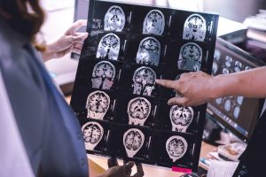
Currently, diagnosing Alzheimer’s relies largely on documentation of any mental decline. However, when diagnosing Alzheimer’s in this way, it has already caused severe brain damage in individuals. What if we could diagnose Alzheimer’s before symptoms started? It is increasingly clear that by the time a typical AD patient comes to diagnosis atrophy is well established. The hope is, future treatments could then target the disease in its earliest stages, before irreversible brain damage or mental decline has occurred.
Neuroimaging is among the most promising areas of research focused on early detection. Today, a standard workup for Alzheimer’s disease often includes structural imaging with magnetic resonance imaging (MRI) or computed tomography (CT). These tests are currently used chiefly to rule out other conditions that may cause symptoms similar to Alzheimer’s but require different treatment. Structural imaging can reveal tumors, evidence of small or large strokes, damage from severe head trauma or a buildup of fluid in the brain.
 There has been a transformation in the part played by neuroimaging in Alzheimer disease (AD) research and practice in the last decades. Diagnostically, imaging has moved from a minor exclusionary role to a central position. In research, imaging is helping address many of the scientific questions and provides insights into the effects of AD and its temporal and spatial evolution. Furthermore, imaging is an established tool in drug discovery, increasingly required in therapeutic trials as part of inclusion criteria, as a safety marker, and as an outcome measure.
In November of 2017, scientists at the University of Twente in The Netherlands, used ‘Ramen’ optical technology in their ongoing research of Alzheimer’s. Using this technology, they can now produce images of brain tissue that is affected by the disease. These images also include the surrounding areas, that may already be showing changes.
Alzheimer’s disease is associated with areas of high protein concentration in brain tissue: plaques and tangles. Raman imaging is now used to get sharp images of these affected areas. It is an attractive technique, because it shows more than the specific proteins involved. The presence of water and lipids, influenced by protein presence, can also be detected.
Compared to MRI, PET and CT imaging, Raman is able to detect areas, smaller than cells, with very high precision. In this way, it can be a very valuable extra technique. The Raman images now show protein activity at neural cell level, but the sensitivity is high enough for detecting areas that are even smaller — as is the case with the brain sample of the healthy person.
No one imaging modality can serve all purposes as each has unique strengths and weaknesses. However, you can trust that Universe Optics designs and manufactures precision lenses for the highest degree of accuracy for any form of neuroimaging systems for diagnosing Alzheimer’s.
Researchers hope to discover an easy and accurate way to detect Alzheimer’s before these devastating symptoms begin. The challenge for the future will be to combine imaging biomarkers to most efficiently facilitate diagnosis, disease staging, and, most importantly, development of effective disease-modifying therapies.