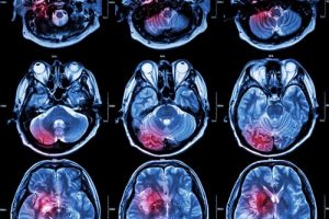
Neuroimaging or brain imaging is the use of various techniques to either directly or indirectly image the structure, function/pharmacology of the nervous system. It is a relatively new discipline within medicine, neuroscience, and psychology. Physicians who specialize in the performance and interpretation of neuroimaging in the clinical setting are neuroradiologists.
Neuroimaging falls into two broad categories:
- Structural imaging, which deals with the structure of the nervous system and the diagnosis of gross (large scale) intracranial disease (such as tumor), and injury, and
- Functional imaging, which is used to diagnose metabolic diseases and lesions on a finer scale (such as Alzheimer’s disease) and also for neurological and cognitive psychology research and building brain-computer interfaces.
Functional imaging enables, for example, the processing of information by centers in the brain to be visualized directly. Such processing causes the involved area of the brain to increase metabolism and “light up” on the scan.
Nervous systems are complex. After all, everything that anyone thinks or does is controlled by its nervous system. To better understand how complex nervous systems work, researchers have used an expanding array of ever more sophisticated tools that allow them to actually see what’s going on. In some cases, neuroscience researchers have had to create entirely new tools to advance their work. With the creation of newly designed, complex tools, guaranteeing clear, precise imaging requires a lens manufactured to the exact specifications. At UKA, we not only design, but, use cutting edge technology to manufacture your specific lens.
In a partnership melding neuroscience and electrical engineering, researchers from UNC-Chapel Hill and NC State University have developed a new technology that will allow neuroscientists to capture images of the brain almost 10 times larger than previously possible – helping them better understand the behavior of neurons in the brain.
A research team made of up of Jeff Stirman, Ikuko Smith and Spencer Smith from UNC-Chapel Hill, wanted to look at activity in neurons across multiple areas at the same time.
To do that, the researchers used a two-photon microscope, which images fluorescence. In this case, it could be used to see which neurons “light up” when active.
Currently, conventional two-photon microscopy systems only looks at approximately one square millimeter of brain tissue at a time. That made it hard to simultaneously capture neuron activity in different areas.
An assistant professor of electrical and computer engineering at NC State, Michael Kudenov’s area of expertise is remote imaging. His work focuses on developing new instruments and sensors to improve the performance of technologies used in everything from biomedical imaging to agricultural research.
After being contacted by the UNC researchers, Kudenov designed a series of new lenses for the microscope. Stirman further refined the designs and incorporated them into an overall two-photon imaging system that allowed the researchers to scan much larger areas of the brain. Instead of capturing images covering one square millimeter of the brain, they could capture images covering more than 9.5 square millimeters.
This advance allows them to simultaneously scan widely separated populations of neurons.
As the group notes in its Nature Biotechnology paper, this work addresses “a major barrier to progress in two-photon imaging of neuronal activity: the limited field of view.”
Universe Kogaku designs and manufactures optical lenses for Two-Photon Imaging systems, security, high tech and electronic applications. We stock 1000’s of standard lens assemblies and can custom design a solution for scanners, CCTV, CCD/CMOS, medical imaging, surveillance systems, machine vision and night vision systems.