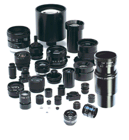
High resolution lenses for machine vision — standard and custom lens design
Image Sensors and the Updated Petri Dish
High Resolution Lenses for machine vision, instrumentation, inspection and vibration-sensitive applications. Standard and custom hi-res lens assemblies.

Image Sensors
The petri dish is likely one of the first “scientific objects” many of us were introduced to in the high school laboratory. Since its invention in the 1800s, the humble petri dish has remained largely unchanged, it went from being made of glass to being made of plastic and rings were added to aid in the stacking process. There had not been any upgrades, until recently when California Institute of Technology moved the petri dish into the 21st century when they incorporated an image sensor.
The image sensor is similar to the ones mobile telephones are equipped with; the installation of the sensors means scientists may not need to be peering at the items in the petri dish through bulky microscopes. Currently, petri dishes are removed from the incubator and studied under a microscope lens; the new ePetri dish, as its been named, lets the scientist or researcher study the organisms without being removed from the incubator. The sensor from the petri dish transmits the data from the dish to a computer at the push of a button.
How it works: With the new technology, a culture is put on the image-sensor chip within the petri dish and the LED display from the sensor scans the image using its scanning light source to transmit data. Once the petri dish is put into the incubator, there is a wire that runs from the chip to a computer which “snaps” pictures of the culture and sends it to the computer screen.
With the new technology, images are saved and transmitted in real time, allowing the scientists the ability to watch the cultures grow in real time. Researchers are embracing this technology because of its ability to image confluent cells, those that grow in close proximity to one another; prior to the new technology it was a very labor-intensive process.
According to a lead author of the study, and a graduate student at Caltech, “the ePetri dish is a small, lens-free microscopy imaging platform” that allows researchers to “streamline and improve cell culture experiments by reducing the risk of contamination and human labor.”
Because some cells behave differently within different parts of a petri dish, such as embryonic stem cells, the new ePetri system reduces the lens limitations of a conventional microscope. With the ePetri system, the lens allows for focus on the entire dish rather than specific regions as items such as embryonic stem cells react differently with other cells in various parts of the petri dish. The ePetri dish technology allows the scientists to follow the cells as they interact across the spectrum of the dish.
The future: The full scope of the ePetri dish’s technology have not been tapped yet, as researchers believe it can be incorporated into other devices such as those that provide microscope imaging capabilities and in medical imaging procedures.
For more information you can contact the sales department at Universe Optics to discuss how the image sensor can assist in medical imaging procedures.