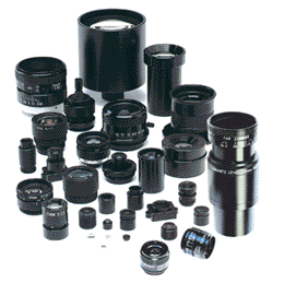
High resolution lenses for machine vision — standard and custom lens design
Imaging Device Isolates Malignant Brain Tumors
High Resolution Lenses for machine vision, instrumentation, inspection and vibration-sensitive applications. Standard and custom hi-res lens assemblies.

Imaging Device
A compact, low price point imaging device has been developed by researchers at the Cedars Sinai Institute and Department of Neurosurgery that has the capability to distinguish malignant brain tumors and other cancerous cells from non-cancerous, healthy tissue and cells.
The camera has a “targeted imaging agent” that is based on a synthetic version of a peptide (small protein) taken from the venom of a death-stalker scorpion. The peptide can focus in on brain tumor cells and when stimulated with a laser in the near infrared portion of the spectrum, a glow is emitted that, while invisible to the naked eye, can be captured on an imaging device.
Studies conducted on laboratory mice that were implanted with human brain tumors allowed researchers to see clear delineations between the tissue with the tumor and the healthy tissue. The near infrared light was able to penetrate into the tissue and identify tumors that would normally have escaped detection.
A research scientist in the study noted that the “tumor-imaging process consists of two parts: deploying a fluorescent ‘dye’ that sticks only to cancer cells, and using a laser and a special camera to make an invisible image visible.” The dye is linked to the peptide and adheres to the malignant cells, completely bypassing the normal, healthy cells. The dye and the peptide are delivered intravenously and the peptide seeks out the tumor. Once the near infrared laser is shined on the tissue, the tumor gives off a glow that is only visible on the imaging device.
In other experimental systems, using different tumor-targeting methods the cameras and systems were too bulky and cost prohibitive. With the new system and its single camera device that can simultaneously take near infrared and white light images the image is captured by alternately strobing the lights on the impacted area. While the human eye can see the normal light, the camera captures both and then superimposes the two images, making it visible to the researchers. The image captured shows the tumor and the white light shows researchers the visible landscape that allows them to see the tumor in context.
The technology is still in the prototype stage with researchers working to make the next generation camera lighter, smaller and more portable allowing it to be easily used in an operating room setting.
Universe Optics is a manufacturer of standard and custom lens assemblies for medical imaging and diagnostic cameras.