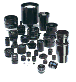
High resolution lenses for machine vision — standard and custom lens design
Descriptions Of Various Types Of Microscopes
High Resolution Lenses for machine vision, instrumentation, inspection and vibration-sensitive applications. Standard and custom hi-res lens assemblies.

Types Of Microscopes
Those who work in and around microscopes on a regular basis understand the myriad types and complexities of microscopes on the market. For those who aren’t intimately involved in the industry, we provide this primer on the various types and classifications of microscopes. Be aware though, that some microscopes and their technologies, fit into more than one category.
Microscope basics:
- Compound microscopes are made up of two lens systems and have a maximum useful magnification of about 1,000x. The two lenses are the objective and the ocular (eye piece).
- A confocal laser scanning microscope differs from compound and stereo microscopes in that these devices are typically used in research facilities. They can scan samples in depth and computer software assembles the data in order to create a 3D image.
- Electron microscopes can magnify up to two million times because of the high energy electrons they create are extremely small. The high energy electrons can take a toll on the sample being observed and this may mean it could lead to the dehydration of the specimen. Biological samples being used with an electron microscope are typically coated with a thin layer of metal prior to being observed.
- A neutron microscope is one that is still in the experimental stage but offers the promise of high resolution and higher contrast than other models.
- An optical microscope is capable of using the visible light (UV light) to capture an image. Optical lenses refract the light. These were the first microscopes invented. Optical microscopes are also found in many other categories in one form or another.
- In the scanning microscope field there are several types: scanning acoustic microscope (SAM); scanning helium ion (SHIM or HeIM) and scanning electron (SEM). With the SAM, the device focuses sound waves to generate an image. SAMs are used to detect tensions or small cracks in objects or to test the elasticity in a biological structure; the SHIM or HeIM uses a beam of helium ions to generate the image. In the field, this is a relatively new technology, having been around for only seven years; SEM utilizes an electron beam that bounces off the sample, they do not go through it. This allows for the capture of a three dimension image.
- With the use of a scanning probe microscope being able to see individual atoms is the strength of this technology. The atom’s image is computer generated.
- A stereo, or dissecting, microscope can magnify up to a maximum of 100x and can offer researchers a 3D image of the specimen. These are typically used to view opaque specimens.
- A TEM, transmission electron microscope, uses an electron beam, which passes through the specimen, to capture a two dimensional image.
- X-ray microscopes use a small wavelength to capture an image. The image resolution with x-ray microscopes is higher than optical models. These devices can observe living microorganisms.
The type of microscope used is predicated on the image needing to be gathered because different microscopes capture and enhance different physical characteristics.