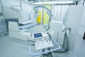
Optical imaging is a technique for non-invasively looking inside the body, as is done with x-rays. But, unlike x-rays, which use ionizing radiation, optical imaging uses visible light and the special properties of photons to obtain detailed images of organs and tissues as well as smaller structures including cells and even molecules. Because it is much safer for patients, and significantly faster, optical imaging can be used for lengthy and repeated procedures over time to monitor the progression of disease or the results of treatment.
 According to the Centers for Disease Control, there is an average of 1.5 million reported cases of Traumatic Brain Injury (TBI) per year resulting in over 1 million emergency department visits, 235,000 hospitalizations and 50,000 deaths annually. Traumatic brain injury is a major public health concern and is characterized by both apoptotic and necrotic cell death in the lesion.
One of the limitations of TBI research is the inability to conduct longitudinal studies either in vitro or in vivo making it difficult to study either the mechanisms or the treatment of TBI over the progression of the disease. Anatomical imaging is usually used to assess traumatic brain injuries and there is a need for imaging modalities that provide complementary cellular information.
Optical imaging allows doctors and researchers to literally see the invisible. At UKA, we design cutting edge lenses to meet the ever-growing demands of optical imaging devices. Our in-house service provides you the top of the line quality from design through manufacturing.
In relationship to blast-Traumatic Brain Injury, Jonathan Estrada, a doctoral student in the School of Engineering at Brown University (Providence, RI), and colleagues are using an optical imaging method to explore the mechanics of cavitation-induced injury to better understand bTBIs.
Estrada is working under the guidance of Christian Franck, along with colleagues from Brown University and the University of Michigan (Ann Arbor, MI). The research team is using a laser, an optical microscope, and rat neurons inside a gel-like substance to mimic brain tissue to examine bTBIs. The laser pulse is sent through the “brain tissue” under the microscope while a high-speed camera—recording 270,000 frames/s—captures the laser creating a bubble, the bubble breaking, and the damage this causes to the rat neurons. The team imaged affected neurons before and immediately after injury, Estrada explains.
The significance of the research team’s work is that while postmortem studies have begun to show differences in brain pathology—such as astroglial (star-shaped glial cell) scarring—between patients exposed to blast injury and those with bTBI, the manifestation of injury over time still isn’t well understood. “Our work, using the simplified bubble and neuron culture model, aims to begin bridging the gap between the mechanics of blast injury and cell damage,” Estrada says.
More research into the specifics of TBI is clearly needed, yet with the precision of optical imaging, the ability to understand the effects on the brain and the long-term progression of the disease.
Universe Kogaku designs and manufactures optical lenses for optical imaging equipment, security, high tech and electronic applications. We stock 1000’s of standard lens assemblies and can custom design a solution for scanners, CCTV, CCD/CMOS, medical imaging, surveillance systems, machine vision and night vision systems.