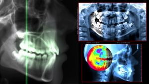
Cone-beam imaging CT (CBCT) scanners offer the advantages of being compact in size, less costly, and more portable than multidetector CT systems. Image quality, however, is often impacted by artifacts such as streaking or shading, associated with cone-beam effects and/or nonuniformity in image quality near the ends of the field-of-view (FOV).
 Cone beam computed tomography (or CBCT, also referred to as C-arm CT, cone beam volume CT, or flat panel CT) is a medical imaging technique consisting of X-ray computed tomography where the X-rays are divergent, forming a cone.
CBCT has become increasingly important in treatment planning and diagnosis in implant dentistry, ENT, orthopedics, and interventional radiology (IR), among other things. Perhaps because of the increased access to such technology, CBCT scanners are now finding many uses in dentistry, such as in the fields of oral surgery, endodontics and orthodontics. Integrated CBCT is also an important tool for patient positioning and verification in image-guided radiation therapy (IGRT).
Research is now underway to improve CBCT image quality. Biomedical engineers at Johns Hopkins University are collaborating with the radiology team at Carestream Health to develop a multi-source CBCT scanner dedicated for extremity imaging. The team has demonstrated that their prototype research scanner reduced cone-beam artefacts over a single-source CBCT scanner and provided a larger longitudinal FOV.
The team has now demonstrated that the prototype research scanner reduced cone-beam artefacts over a single-source CBCT scanner and provided a larger longitudinal FOV.
The scanner is a c-shaped, self-shielded carbon fiber hull enclosing the source and detector, enabling an arm or leg to be placed through a door within the “C”. The inner diameter of the gantry is 20 cm, and includes an expanded region for placement of a foot. The circular detector orbit extends to 210°, with the flat-panel X-ray detector (FPD) passing within the closed door during a scan. The source-to-axis distance is 40.4 cm, and the source-to-detector distance is 53.8 cm.
The scanner’s X-ray source consists of three separate anode-cathode units evenly distributed with 12.7 spacing along the longitudinal direction. Each anode-cathode axis is oriented along the longitudinal axes and angulated to present a focal spot at the center of the detector. Each source has a separate primary collimator with three output windows on the X-ray tube housing, allowing each source to cover a large FOV at the FPD. The FPD was read in 2 x 2 binning mode at a pixel size of 0.278 mm, with a nominal readout rate of 25 frames per second.
The researchers investigated the image quality and dose characteristics of the prototype scanner. For the study, they acquired images from both single-source and three-source configurations. The scan protocol for all acquisitions used 90 kV, 6 mA tube current and 20 ms pulse width for each source. A total of 600 projections (200 for each source) were acquired. They utilized a custom filtered back projection (FBP) algorithm for reconstruction, using a simple linear combination of the three single-source FBP reconstructions via a voxel-based weighting “map”. They also used a soft-tissue kernel to visualize soft-tissue details of muscle, cartilage, tendons and ligaments above the knee and joint space.
With the priority of this research being image quality, it’s imperative that the lens used in the cone beam scanners offer precision, accuracy, and clarity. Universe Optics is dedicated to designing and manufacturing precision lenses to be used in this field of technology.
Sampling characteristics of the three-source configuration were superior overall. Areas of greatest reduction in noise and cone-beam artefacts were identified near the central plane of each X-ray source, increasing at locations intermediate to each source. The authors reported that this configuration demonstrated a more complete 3D sampling with an overall reduction in the “null cone” of unsampled frequencies.
“The multiple-source configuration appears particularly beneficial for imaging scenarios in which the anatomy contains high-contrast surfaces at large distance from the central axial plane and/or throughout the FOV, as with the hand, knee or foot,” wrote the authors.
One of the major benefits of cone-beam imaging is that they can produce the same kind of high-quality images as a CT (CAT) scan with less radiation. Imaging using this technology will continue to improve, and we, at UKA, are on the cutting edge of specific lenses designed for such equipment.